Formulation and evaluation of silver nanoparticles as antibacterial and antifungal agents with a minimal cytotoxic effect
Keywords:
Silver nanoparticles, antibacterial, antifungal, cytotoxicity, micro-plate assay, release kineticsAbstract
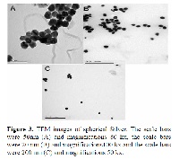
Preparation of non-biodegradable nonoparticles is a fast growing field, which is vital in both nanomedicine and nanotechnology applications. In this investigation, our attention will be focused on the preparation and evaluation of colloidal silver nanoparticles as antibacterial and antifungal agents. The colloidal silver nanoparticles have been prepared employing standard chemical reduction methods. The colloidal silver nanoparticles were characterized using transmission electron microscopy TEM, zeta potential, photo correlation spectroscopy PCS, and in vitro release kinetics. The particles thus obtained were spherical in shape and having an average particles size of 5-20 nm , zeta potentials of -25.5 to -38.3 mV, and the release kinetics was following zero order kinetics with r2>0.96. The dissolution data indicates that the release of the silver nanoparticles is inversely correlated with the size of the nanoparticles i.e. the release increased with smaller particles. The results suggest that the Ag NPs would be stable in the pharmaceutical preparations and will be easily to the infection site. The colloidal silver nanoparticles were found to be very efficient antibacterial agents for different types of bacteria. The bacteria studied were namely: E. coli, S. coccus, Salmonellae, and P. aeruginosa. The associated antifungal effects were also investigated for Aspergillus and Pencillium. . Cytotoxicity of the nanoparticle was studied using human fibroblast cell line. It was concluded that cytotoxicity is concentrations dependant. The results provided strong evidence that could warrant the consideration of silver nanoparticles as antibacterial and antifungal agent that could circumvent the side and passive effects of the conventional antibiotics.
References
Alivisatos AP, Semiconductor Clusters,
Nanocrystals and Quantum Dots. Science
;271:933-937.
Elechiguerra JL, Burt J, Morones JR,
Camacho-Bragado A, Gao X, Lara HH,
Yacaman MJ. Interaction of Silver
Nanoparticles with HIV-1. J.
Nanobiotechnol. 2005;3:1–10.
Feng QL, Wu J, Chen GQ, Cui FZ, Kim TN,
Kim JO, Biomed. Mater J. Res.
;52:662.
Petica A, et al. Colloidal silver solutions with
antimicrobial properties', Materials Science
and Engineering: B. 2008;152(1-3):22-27.
Israelachvili JN, Intermolecular Surface
Forces, 2nd ed, Academic Press, San Diego,
Fendler JH, Korean J. Colloidal
Nanoparticles and Nanoparticulate Films
Grown at the Air-Water Interface in
Reactions and Synthesis in Surfactant
Systems Chem. Eng. 2001;18(1):1–13.
Henglein A, Meisel D. Spectrophotometric
Observations of the Adsorption of
Organosulfur Compounds on Colloidal
Silver Nanoparticles J. Phys. Chem. B
;102: 8364.
Kreibig U, Vollmer M. Optical Properties of
Metal Clusters. Springer: New York, 1995.
El-Sayed MA. Some Interesting Properties
of Metals Confined in Time and Nanometer
Space of Different Shapes. Acc. Chem. Res.
;34:257-264.
Frens G. Controlled Nucleation for the
Regulation of the Particle Size in
Monodisperse Gold Suspensions Nature
Phys. Sci. 1973;241:20-22.
Christian GD, Feldman FJ. Atomic
Absorption Spectroscopy Applications in
Agriculture, Biology and Medicine, John
Wiley & Sons, Inc., 1970.
Valcarcel M, Luque de Castro M D, `FlowInjection Analysis principles an
applications', Ellis Horwood Ltd., Halsted
Press a division of John Wiley & Sons, 1987.
Thompson M, Walsh JN. Handbook of
Inductively Coupled Plasma Spectrometry',
nd ed., Blackie & Son Ltd., 1989.
Tenover F, et al. Comparison of traditional
and molecular methods of typing isolates of
Staphylococcus aureus. Journal of Clinical
Microbiology, 1994;32(2):407.
Uznanski P, et al, Oxidation of photochromic
spirooxazines by coinage metal cations. Part
I. Reaction with AgNO3: formation and
characterisation of silver particles. New
Journal of Chemistry, 2001. 25(12): 1486-
Choi O and Hu Z. Size dependent and
reactive oxygen species related nanosilver
toxicity to nitrifying bacteria. Environmental
science & technology, 2008;42(12):4583-
Fu J, et al. Construction of antibacterial
multilayer films containing nanosilver via
layer by layer assembly of heparin and
chitosan silver ions complex. Journal of
Biomedical Materials Research Part A,
;79(3):665-674.
International Standard Methods for
determination of particle size distribution
part 8: photon correlation spectroscopy,
International organization for standardization
ISO, 13321, 1996.
Riddick T. Control of colloid stability
through zeta potential: with a closing chapter
on its relationship to cardiovascular disease:
Published for Zeta-Meter, inc., by Livingston
Pub Co., 1968.
Margalit R, Okon M, Yerushalmi Y, Avidor
E. Bioadhesive Liposomes for Topical Drug
Delivery: Molecular and Cellular Studies. J.
Controlled Release 1992;19:275-287.
Yerushalmi N, Margalit R. Physicochemical
Evaluation of a Stability-Driven Approach to
Drug Entrapment in Regular and in SurfaceModified Liposomes. Biochem. Biophys.
Acta.1994; 1189:13-20.
Margalit R, Alon R, Linenberg M, Rubin I,
Roseman TJ, Wood RW. Liposomal Drug
Delivery: Thermodynamic and Chemical
Kinetic Considerations J. Controlled Release
;17:285-296.
Margalit R, Okon M, Yerushalmi N and
Avidor E. Liposome-encapsulated silver
sulfadiazine (SSD) for the topical treatment
of infected burns: Thermodynamics of drug
encapsulation and kinetics of drug release J.
Controlled Release 1992;19:275.
Yerushalmi N and Margalit R.
Physicochemical Evaluation of a StabilityDriven Approach to Drug Entrapment in
Regular and in Surface-Modified Liposomes,
Biochem. Biophys. Acta 1994;1189:13.
Margalit R, Alon R, Linenberg M, Rubin RJ
Roseman RW, Wood J. Liposomal drug
delivery: thermodynamic and chemical
kinetic considerations, J. Controlled Release
;17:285.
Kim J, et al, Antimicrobial effects of silver
nanoparticles. Nanomedicine:
Nanotechnology, Biology and Medicine,
;3(1):95-101.
Sondi I and Salopek-Sondi B. Silver
nanoparticles as antimicrobial agent: a case
study on E. coli as a model for Gramnegative bacteria. Journal of Colloid and
Interface Science, 2004;275(1):177-182.
Gill S, et al, Insights on evolution of
virulence and resistance from the complete
genome analysis of an early methicillinresistant Staphylococcus aureus strain and a
biofilm-producing methicillin-resistant
Staphylococcus epidermidis strain. Journal
of bacteriology, 2005;187(7):2426.
Choi O, Hu Z. Size dependent and reactive
oxygen species related nanosilver toxicity to
nitrifying bacteria. Environmental science &
technology, 2008;42(12):4583-4588.
Singh N, et al, NanoGenotoxicology: The
DNA damaging potential of engineered
nanomaterials. Biomaterials, 2009;30(23-
:3891-3914.
Samberg M, Orndorff P, Monteiro-Riviere
N. Antibacterial efficacy of silver
nanoparticles of different sizes, surface
conditions and synthesis methods.
Nanotoxicology, (0):1-10.
Pal S, Tak Y, Song J. Does the antibacterial
activity of silver nanoparticles depend on the
shape of the nanoparticle? A study of the
gram-negative bacterium Escherichia coli.
Applied and environmental microbiology,
;73(6):1712.
Li X, et al, Effects of molecular weight and
concentration of chitosan on antifungal
activity against Aspergillus Niger. Iran
Polym J. 2008;17:843-852.
Ma Y, Zhou T, and Zhao C. Preparation of
chitosan-nylon-6 blended membranes
containing silver ions as antibacterial
materials. Carbohydrate research,
;343(2):230-237.
Berger T, et al, Antifungal properties of
electrically generated metallic ions.
Antimicrobial Agents and Chemotherapy,
;10(5):856.
Chen Y, et al, Anti-bacterial and cytotoxic
properties of plasma sprayed silvercontaining HA coatings. Journal of Materials
Science: Materials in Medicine,
;19(12):3603-3609.
Sung W, Park K, Lee D. Poster Session:
Chemical Genomics and Drug Discovery;
Antifungal effects and its mode of action of
silver nanoparticles against human
pathogenic fungi. 2007;2007:355.
Nierman W, et al, Genomic sequence of the
pathogenic and allergenic filamentous
fungus Aspergillus fumigatus. Nature,
;438(7071):1151-1156.
Suh J, Wirtz D, Hanes J. Efficient active
transport of gene nanocarriers to the cell
nucleus. Proceedings of the National
Academy of Sciences of the United States of
America, 2003;100(7):3878.